01
Collect auricular cartilage
Three tiny 2.5 mm punch samples are taken from the bowl of the ear under local anaesthetic.

Mytocel MSK · Standalone autologous micrograft treatment
Mytocel MSK uses a few tiny samples of auricular cartilage from the patient’s ear. They are mechanically prepared at the point of care into an autologous micrograft suspension, then delivered to the affected joint as the treatment itself.
The standalone Mytocel MSK pathway
Mytocel MSK is a complete treatment route in its own right. Auricular cartilage is collected, mechanically prepared and returned to the clinically selected joint during one appointment, without laboratory culture.
Three tiny 2.5 mm punch samples are taken from the bowl of the ear under local anaesthetic.
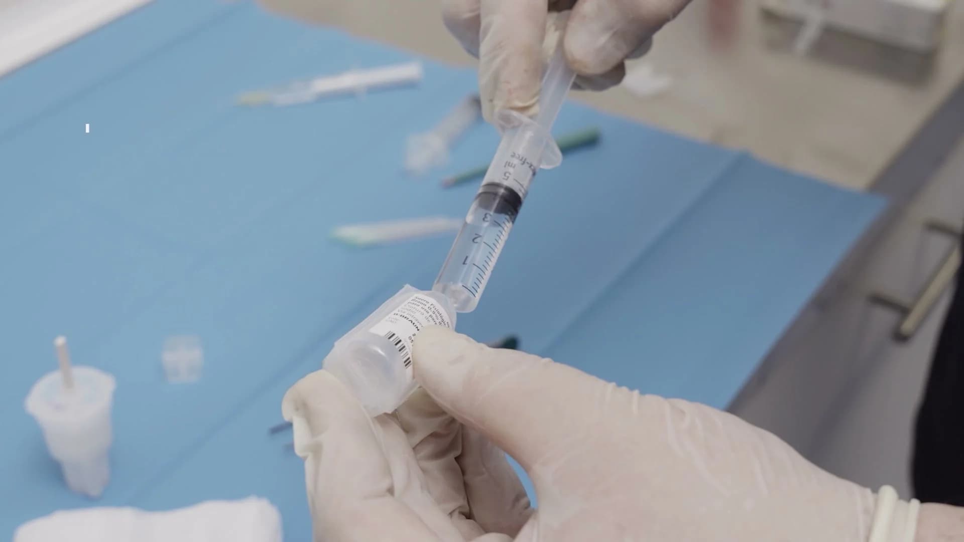
Cartilage and saline pass through a closed, single-use cartridge that creates microscopic autologous fragments.
Approximately 4 ml of freshly prepared suspension retains tissue-derived cells and elements of their native matrix.
The freshly prepared micrograft suspension is injected into the affected joint in the same session.
What the technology changes
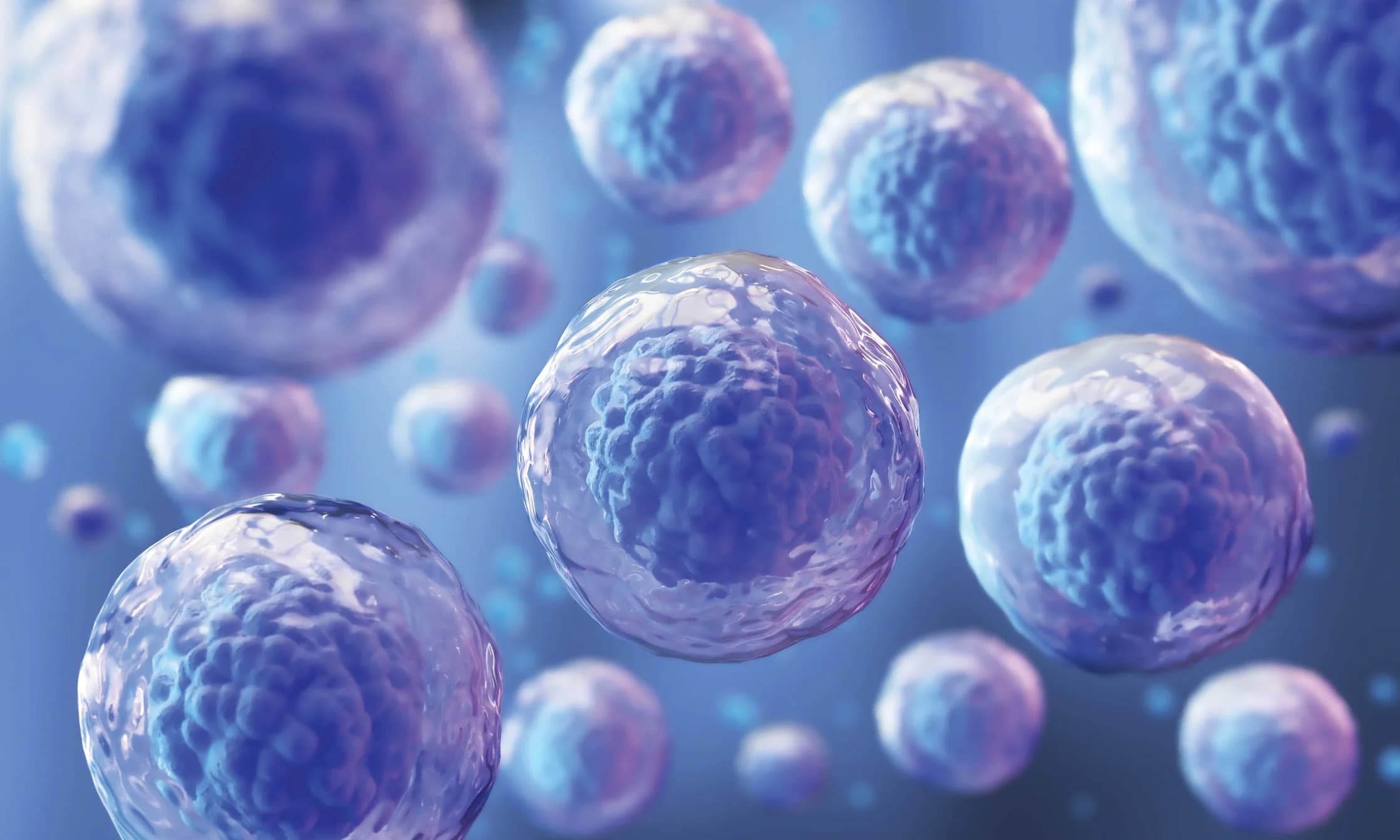
Autologous Micrografting Technology uses controlled mechanical disaggregation. A few tiny samples of auricular cartilage are divided into microscopic fragments while retaining cells and elements of their native extracellular matrix. The resulting AMT suspension is delivered directly to the affected joint in the same appointment.
The practical difference is time and handling. Traditional cell-implantation pathways can require a harvest operation, laboratory culture and a later procedure. Mytocel MSK is designed to prepare autologous biological material in minutes, beside the patient.
Where it may be used
The donor tissue in the standard Mytocel MSK route is auricular cartilage. What changes is the joint or soft-tissue problem being treated, based on examination, imaging and whether the mechanics still make biological treatment credible.
Mild-to-moderate wear where the joint remains worth protecting and a regenerative injection is clinically appropriate.
The knee is a common target, with selected hip, shoulder or ankle problems considered on their individual anatomy and imaging.
Smaller joints may also be considered, including thumb-base osteoarthritis, where treatment can be delivered precisely.
Mytocel MSK may have a role beyond joint cartilage where the diagnosis, tissue quality and loading plan support its use.
What Mytocel MSK avoids
The preparation does not rely on multi-week cell culture, enzymatic digestion or genetic manipulation. It remains the patient’s own tissue, mechanically prepared for same-session use.
What Mytocel MSK delivers
Mechanical preparation creates microscopic fragments that retain tissue-derived cells and elements of their surrounding extracellular matrix. The suspension is then delivered as Mytocel MSK itself, with recovery planned for the joint being treated.
Evidence
Autologous Micrografting Technology has been studied in joint osteoarthritis and other musculoskeletal applications. That evidence belongs to the Mytocel MSK pathway and should be considered separately from evidence for NANO ACi or any other combined procedure. Suitability and likely benefit remain individual clinical questions.
The key points are the auricular-cartilage source, point-of-care preparation and delivery as a standalone same-session treatment.

Read the patient-facing treatment guide, suitability information and Mytocel MSK evidence at London Cartilage Clinic.
Read more

See where standalone Mytocel MSK sits among the wider cartilage options.
Read more

NANO ACi is a separate cartilage technique that uses Mytocel-prepared micrografts as one component of a fuller protocol.
Read more
Prepared at the point of care and delivered in one session
Professor Lee’s assessment determines whether Mytocel MSK is appropriate for the affected joint, based on diagnosis, imaging and the mechanical state of the tissues around it.